Skull Base & Orbit
Skull base compartments and foramina, cranial nerve tracking, cavernous sinus, petrous apex, jugular foramen, orbit and optic pathway disease. 8 topics.
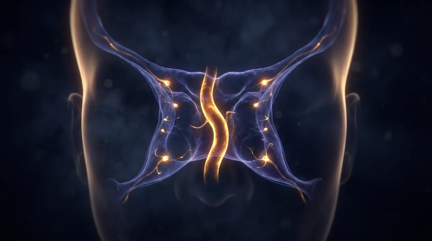
Cavernous Sinus: Who Sits Where
The cavernous sinus is a thin-section, high-resolution, wide-window MR read (CT cannot separate non-enhancing nerves from enhancing venous blood).…
Clivus: Midline Chordoma vs Off-Midline Chondrosarcoma
Triage the clival mass on two facts first — midline vs off-midline, then T2 brightness/matrix — to split chordoma (midline, extremely T2-bright) from…
Complications of Sinusitis: Is It Crossing a Wall?
When you see sinusitis, ask 'is this just sinusitis or is it crossing a wall?' — septum (preseptal vs postseptal), bony table, dura, or vessel wall.…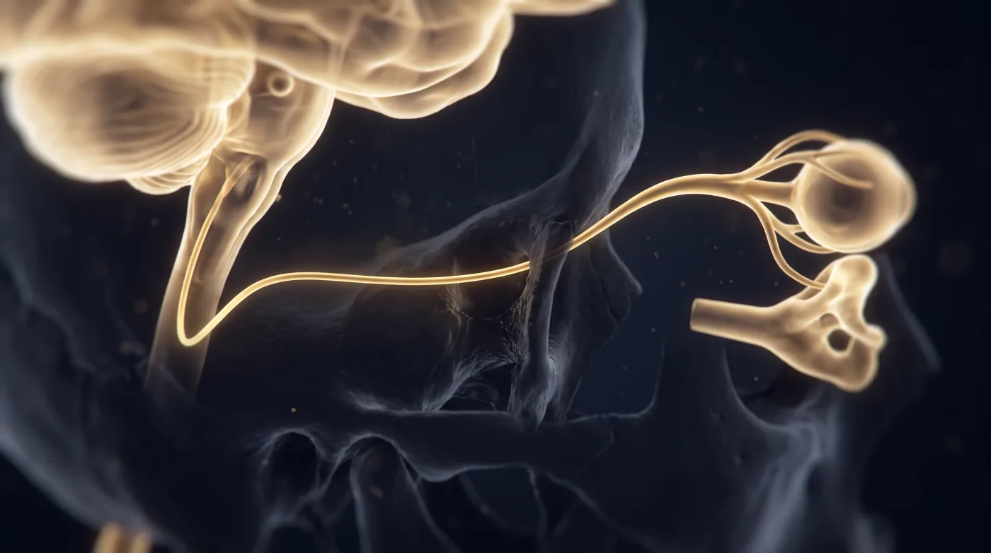
Cranial Nerve Tracking: Follow the Four-Segment Path
Read every cranial nerve as a four-segment journey (brainstem nucleus → cisternal segment → skull-base space/foramen → end organ) and chase the…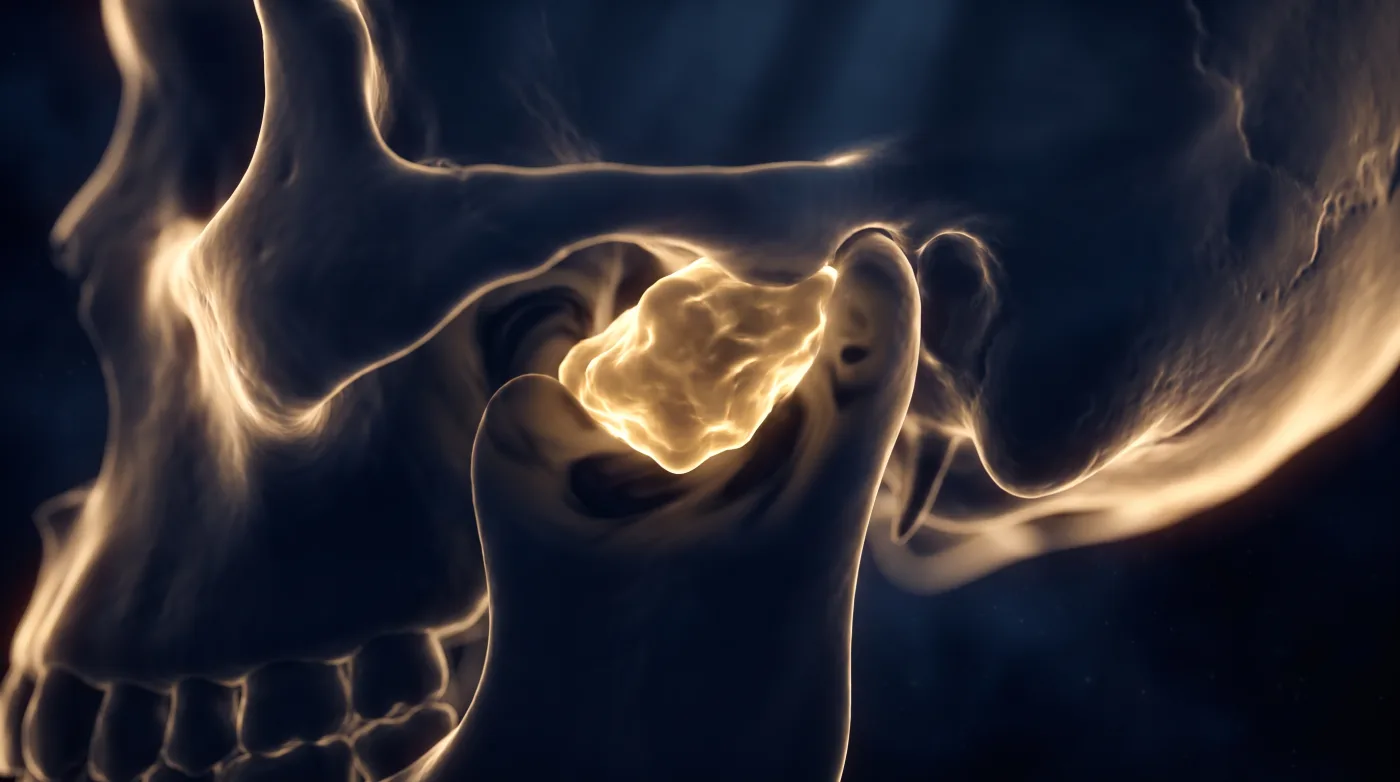
Jugular Foramen: Let the Bone Margin Call It
Close the jugular-foramen differential on two CT features first — the bone margin (destroyed vs scalloped vs sclerotic) and the growth vector — then…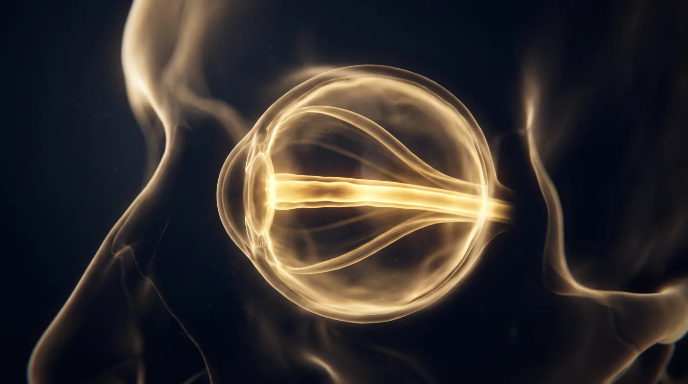
Orbit Anatomy: Septum and Cone First
Localize every orbit case on two anatomic axes before naming anything: the septum (pre- vs post-septal = the infection/triage line) and the cone…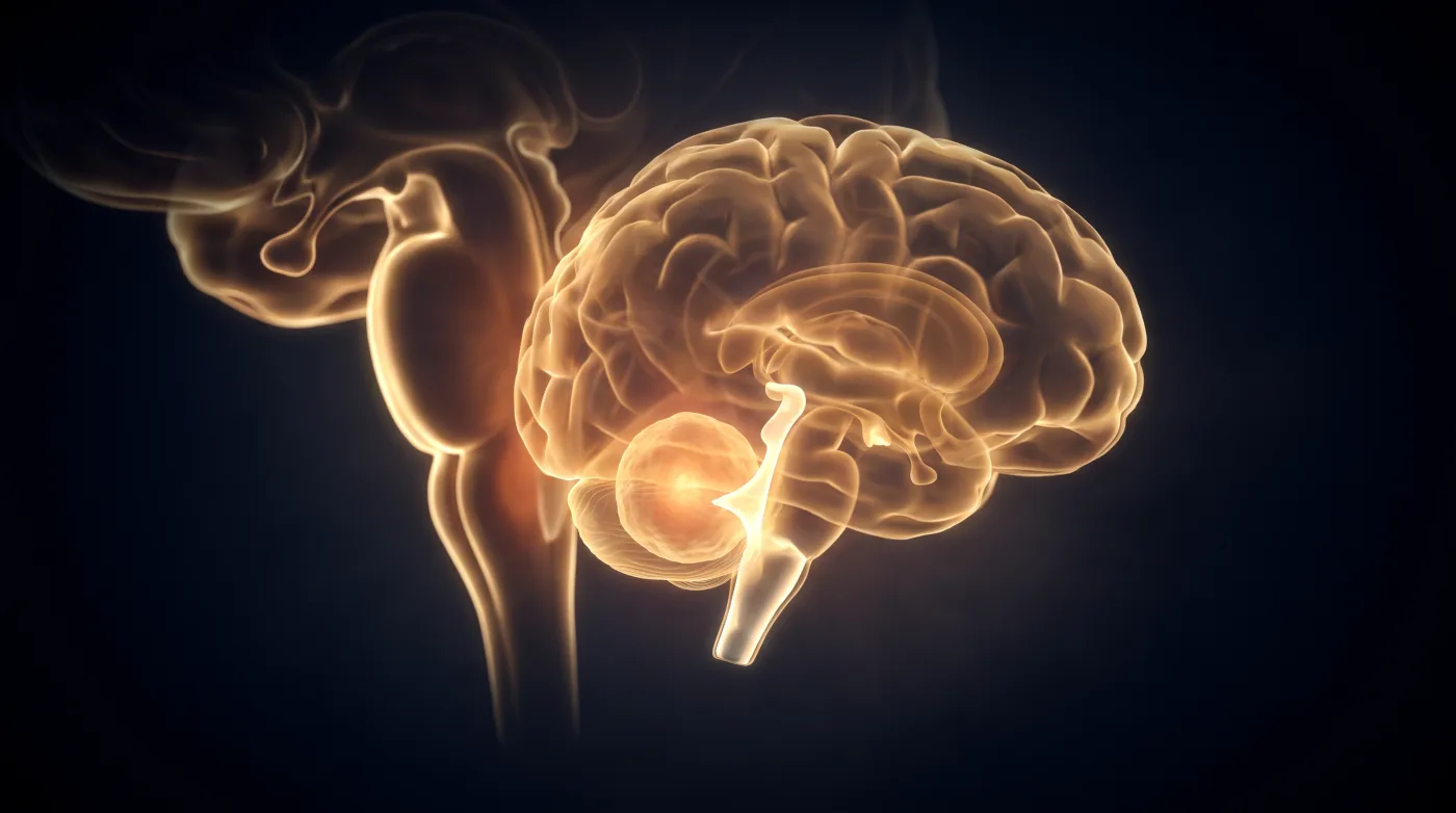
Posterior Fossa Masses: Anchor to the Fourth Ventricle
In the small posterior-fossa box the differential is usually broad, so don't force a single diagnosis — anchor it. Fix a pediatric mass to the fourth…