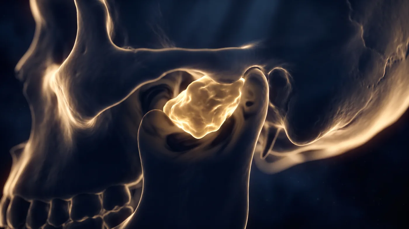
Skull Base & Orbit
Jugular Foramen: Let the Bone Margin Call It
Fellowship-level neuroradiology teaching by E. Brooke Schrickel, MD. Open this topic in the interactive reading room →
Watch · concise explainerVisual summary
Core clinical idea
Close the jugular-foramen differential on two CT features first — the bone margin (destroyed vs scalloped vs sclerotic) and the growth vector — then confirm with enhancement and flow voids. Paraganglioma, schwannoma, and meningioma each have a signature bone behavior; flow voids are the single best binary discriminator.
Bottom line
Read the bone and the vector first — permeative+flow voids = paraganglioma, scalloped = schwannoma, sclerotic = meningioma — then confirm with the denervated muscles.
Core workstation questions
- Is the bone margin destroyed (paraganglioma), scalloped (schwannoma), or sclerotic+permeative (meningioma)?
- Which way is it growing — superolateral, superomedial, or centrifugal?
- Are there flow voids? (Paraganglioma vs not.)
- Is the jugular spine intact?
- Any secondary denervation — vocal cord, constrictors, hemitongue?
- Could this be a metastasis rather than the classic three?
What changes reporting / management
- Lead the report with the bone pattern and vector; they carry the differential before enhancement.
- Call flow voids explicitly — present points to paraganglioma and away from schwannoma/meningioma.
- When uncertain about paraganglioma, recommend or correlate with an octreotide scan (positive in the vast majority).
- Sweep for secondary cranial-nerve denervation (vocal cord/constrictor/hemitongue) to confirm the diagnosis and catch a tumor arising from a neighboring nerve.
- Keep metastasis on the differential for a destructive, diffusely enhancing JF mass that doesn't fit the big three.
Practical traps
- Mistaking a subtle glomus jugulare for a prominent or asymmetric jugular bulb normal variant (true lesion shows T2 hyperintensity, flow voids, avid enhancement).
- Over-weighting the growth vector — a paraganglioma can grow directly superior; the diagnosis rests on permeative bone + flow voids + enhancement.
- Missing that a hypoglossal (CN XII) schwannoma can extend secondarily into the jugular foramen — the denervated hemitongue is the tell.
Teaching pearls
- Flow voids are the tiebreaker — present means paraganglioma.
- Destroyed bone = paraganglioma, scalloped = schwannoma, sclerotic = meningioma.
- A missing jugular spine is permeative destruction until proven otherwise.
- The denervated muscle both confirms the diagnosis and can betray a tumor arising from the neighboring nerve.
Teaching visuals
Source lectures
- Imaging Approach to the Jugular Foramen
Educational material for radiology residents and neuroradiology fellows. Nothing here drives individual patient care, and it contains no patient data.