Skull Base & Orbit
Cranial Nerve Tracking: Follow the Four-Segment Path
Fellowship-level neuroradiology teaching by E. Brooke Schrickel, MD. Open this topic in the interactive reading room →
Core clinical idea
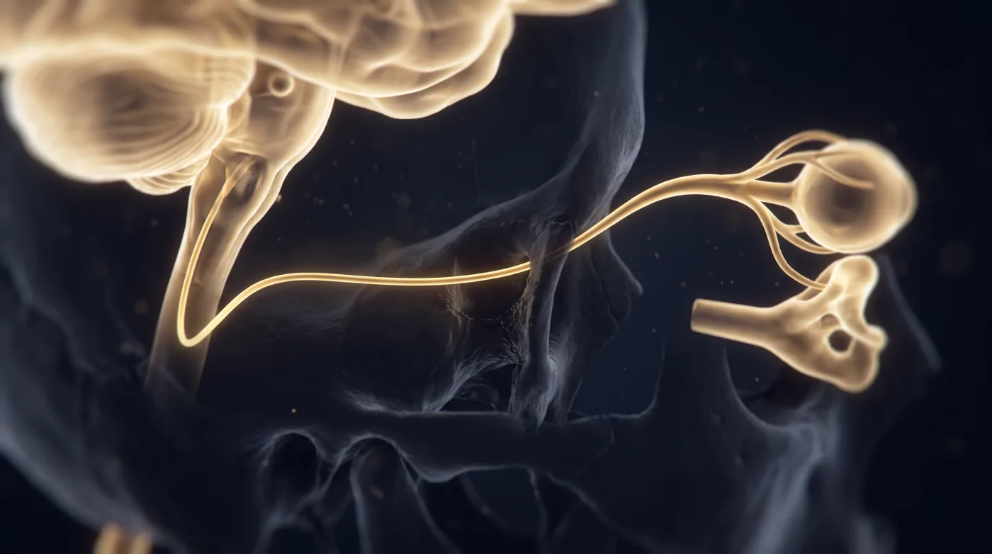
Read every cranial nerve as a four-segment journey (brainstem nucleus → cisternal segment → skull-base space/foramen → end organ) and chase the suspected nerve along its whole path, including the bone it traverses. The miss is the skull-base tumor or end-organ clue, not the nerve itself.
Bottom line
Name the nerve from the history, then track it brainstem-to-end-organ and check the bone it runs through — the miss is the skull-base tumor, not the nerve.
Core workstation questions
- Which nerve does the deficit name, and did I follow its whole course (brainstem to end organ)?
- Did I review the bone/foramen the nerve passes through for a mimicking or compressive lesion?
- Optic nerve: nerve or sheath? Is a normal nerve visible centrally on the coronal?
- CN III palsy: is the pupil involved? (Aneurysm workup vs microvascular.)
- Painful ophthalmoplegia at the cavernous sinus/apex: did I list lymphoma/mets/sarcoid alongside Tolosa-Hunt?
- CN VI palsy: did I trace it up the clivus and inspect the clival bone and sinuses?
- Ambiguous apex/cavernous mass: do the extraocular muscles tell me which nerve?
- CN V disease: have I described full extent for resectability and radiation planning?
What changes reporting / management
- Protocol by nerve group (orbit MR covers II/III/IV/VI; CN V extends lower to the mandible for V3) so the lesion isn't clipped.
- On the optic nerve, use the coronal to call nerve vs sheath, then assign the three-pattern differential (neuritis/glioma vs smooth meningioma vs lumpy sarcoid-mets-lymphoma).
- At first optic neuritis, hunt for a brain white-matter lesion because it changes MS risk stratification and management.
- CN III palsy with a dilated pupil drives urgent CTA/MRI for PComm aneurysm; pupil-sparing suggests microvascular/ischemic.
- Report cavernous sinus/orbital-apex painful ophthalmoplegia as Tolosa-Hunt vs lymphoma/mets/sarcoid, not Tolosa-Hunt alone.
- On any CN VI palsy, trace the nerve up the clivus through Dorello's canal and search the clival bone/sinuses for the culprit lesion.
- Compare extraocular muscles side to side; selective recti atrophy with spared superior oblique/lateral rectus names a CN III process.
- For CN V disease, report precise extent because it dictates resectability and the radiation field.
- Read homogeneous enhancement/filling of Meckel's cave (normally CSF + rootlets) as perineural tumor and follow V2 to the pterygopalatine fossa.
- Call mastication-muscle enhancement + T2 hyperintensity acute denervation (secondary sign), confirmed on non-contrast, not a primary muscle mass.
Practical traps
- Anchoring on a benign nerve read before following the nerve and checking the foramina for perineural tumor spread.
- Calling lumpy optic-sheath disease metastasis in a known-cancer patient when sarcoid looks identical.
- Treating Tolosa-Hunt as a primary diagnosis rather than one of exclusion.
- Mistaking denervation muscle signal/atrophy for a primary muscle mass instead of a sign pointing back up the nerve.
- Missing the V3-to-VII auriculotemporal hop (behind the mandibular ramus) as the route by which a facial/temple skin cancer causes facial weakness.
- Reading Meckel's cave as normal when homogeneous enhancement signals perineural spread.
- Stopping at the cistern — a normal cisternal segment does not exclude a canal/foramen lesion.
- Missing perineural spread along V2/V3 by not following the nerve through rotundum/ovale.
- Overlooking CN IV because of its dorsal midbrain exit into the ambient cistern.
- Not side-comparing, so subtle asymmetric enlargement/enhancement is missed.
Teaching pearls
- Follow the nerve the whole way and read the bone around it — the lesion is usually at the skull base, not on the nerve.
- On the optic nerve, the coronal decides nerve vs sheath: normal nerve in the center = sheath process.
- A dilated pupil with a third-nerve palsy is an aneurysm until the CTA says otherwise.
- Facial weakness or facial pain? Think tumor first.
- When the mass could be any nerve, the atrophic muscle tells you which one.
- CN V's job is extent — resectability and the radiation field depend on how far the tumor has tracked.
- A quiet Meckel's cave is CSF and rootlets; homogeneous enhancement there is perineural tumor.
- Facial weakness with a temple skin cancer? Check behind the mandibular ramus — V3 can hand disease to VII.
- Mastication-muscle enhancement is denervation pointing up the nerve, not a muscle tumor.
- Follow the nerve to its target — the lesion is often in the segment you skipped.
Teaching visuals
Source lectures
- Cranial Nerves I-VI. Part 1
- Cranial Nerves I-VI. Part 2
- Cranial Nerve Tracking (Thin T2)
Educational material for radiology residents and neuroradiology fellows. Nothing here drives individual patient care, and it contains no patient data.