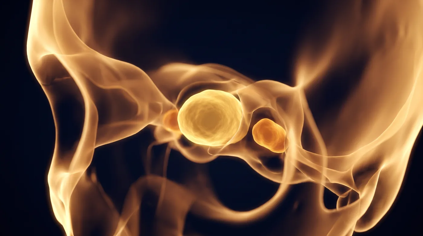
Skull Base & Orbit
Clivus: Midline Chordoma vs Off-Midline Chondrosarcoma
Fellowship-level neuroradiology teaching by E. Brooke Schrickel, MD. Open this topic in the interactive reading room →
Watch · concise explainerVisual summary
Core clinical idea
Triage the clival mass on two facts first — midline vs off-midline, then T2 brightness/matrix — to split chordoma (midline, extremely T2-bright) from chondrosarcoma (off-midline petroclival, rings-and-arcs). Never let an invasive pituitary adenoma or petroclival meningioma slip past on epicenter/bone cues, and recognize skull base osteomyelitis early in a diabetic because that's the diagnosis where speed changes outcome.
Bottom line
Midline vs off-midline and T2 brightness split chordoma from chondrosarcoma — but in a diabetic, the marrow signal is skull base osteomyelitis until proven otherwise.
Core workstation questions
- Midline or off-midline (petroclival)?
- How bright on T2 — extremely (chordoma) or with chondroid dark areas / rings-and-arcs (chondrosarcoma)?
- Is the epicenter actually the sella (invasive pituitary adenoma)?
- Dural tail + hyperostosis (petroclival meningioma)? High CT density (fibrous dysplasia)?
- Diabetic/immunocompromised with marrow signal + mastoid fluid + prevertebral collection — skull base osteomyelitis?
- Is mastoiditis secondary to a nasopharyngeal mass?
What changes reporting / management
- For chordoma, describe soft-tissue extent and vascular encasement (basilar artery, prepontine cistern) for surgical planning.
- Use rings-and-arcs calcification / chondroid matrix, not location alone, to call chondrosarcoma; flag bilateral petroclival chondrosarcoma for Maffucci/enchondromatosis (check hands).
- Identify a sellar epicenter and destroyed sellar floor to catch an invasive pituitary adenoma masquerading as a clival mass.
- Use hyperostosis + dural tail to separate petroclival meningioma from the destructive tumors; correlate high CT density for fibrous dysplasia.
- Escalate skull base osteomyelitis (diabetic, marrow signal, mastoid/middle-ear opacification, prevertebral/retropharyngeal collection, usually Pseudomonas) as an early, high-mortality diagnosis.
Practical traps
- Treating midline-vs-off-midline as a law — chondrosarcoma can be midline; matrix (rings-and-arcs) makes the chondroid call.
- Missing an invasive pituitary macroadenoma presenting as clival pathology by not finding the sellar epicenter.
- Calling simple otitis media/mastoiditis when it is secondary to a nasopharyngeal carcinoma obstructing the eustachian tube.
Teaching pearls
- Midline + extremely T2-bright = chordoma; off-midline + rings-and-arcs = chondrosarcoma — and matrix beats location.
- An invasive pituitary adenoma can masquerade as a clival mass; find the sellar epicenter.
- Petroclival meningioma gives you hyperostosis; chordoma and chondrosarcoma destroy.
- Diabetic + abnormal clival marrow + mastoid fluid + prevertebral collection = skull base osteomyelitis — call it early, it kills.
Teaching visuals
Source lectures
- Imaging of the Clivus
Educational material for radiology residents and neuroradiology fellows. Nothing here drives individual patient care, and it contains no patient data.