Vascular & Stroke
CTA/CTP technique, stenosis and dissection, aneurysms, acute stroke core/penumbra, hemorrhage, vessel wall imaging, PRES/RCVS. 8 topics.
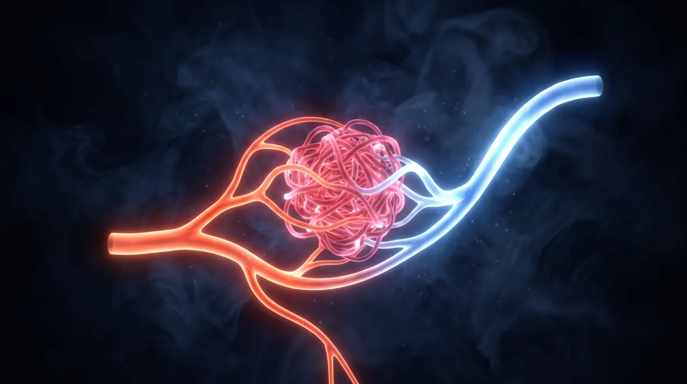
AVM vs Dural AV Fistula: Where the Shunt Lives
Both are arteriovenous shunts, but where the shunt lives and how it drains decide everything: a true (pial) AVM has a parenchymal nidus fed by…
Blunt Cerebrovascular Injury: It Hides — Screen and Grade
Blunt cerebrovascular injury hides: the classic patient has a deficit delayed or out of proportion to the scan because a torn intima emboli before…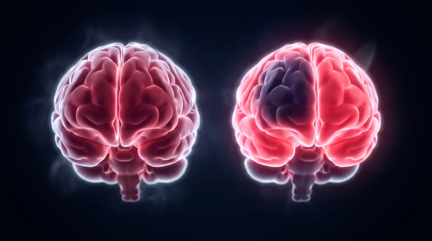
Diamox CT Perfusion: A Stress Test for Flow Reserve
Diamox CTP is a stress test, not an acute-infarct study: image at rest, give the vasodilator, image again, and ask whether the chronically narrowed…
Intracranial Arterial Anatomy: Naming the Segment Is the Report
Naming the segment is the report, not pedantry: the segment, side, and threatened perforators decide clip vs. coil vs. flow-diversion and predict…
Intracranial Hemorrhage: Name the Pattern & the Source
The clinician already knows there's blood — your job is to name the likely source, because each source drives a different treatment. Read it in…
PRES: Reversible — If You Name the Complications
Both are treatable and usually reversible, so the value of the read is the prompt diagnosis plus naming the complications that flip prognosis. PRES…
Perfusion MRI: A Context Modifier, Never a Standalone Read
Perfusion is a context modifier, never a standalone read: triangulate rCBV (and its corrected map), CBF, and transit times against conventional…