Skull Base & Orbit
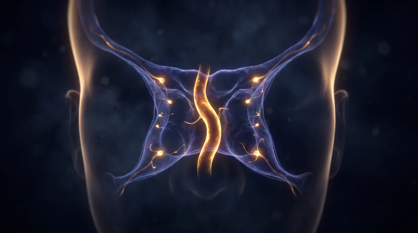
Cavernous Sinus: Who Sits Where
Fellowship-level neuroradiology teaching by E. Brooke Schrickel, MD. Open this topic in the interactive reading room →
Watch · concise explainerVisual summary
Core clinical idea
The cavernous sinus is a thin-section, high-resolution, wide-window MR read (CT cannot separate non-enhancing nerves from enhancing venous blood). Once technique is right, localize against wall anatomy — CN III/IV/V1/V2 ride in the lateral dural wall while VI runs intracavernous next to the carotid (caught early) — and read the sinus as a valveless venous hub for infection spread, thrombosis, and carotid-cavernous fistula.
Bottom line
Cavernous sinus is a thin-section MR read against wall anatomy — wall nerves III/IV/V1/V2 vs intracavernous VI and carotid — and a valveless venous hub when you're chasing fistula or thrombosis.
Core workstation questions
- Is this actually a thin-section, wide-window MR — or am I being asked to clear the cavernous sinus on CT?
- Lateral wall (III/IV/V1/V2) or intracavernous (VI + carotid)?
- Is the medial wall deficient or are the carotids 'kissing' — a transsphenoidal hazard?
- Suspecting CCF/thrombosis: did I follow the valveless drainage map (ophthalmic veins, petrosal sinuses, intercavernous cross-flow)?
- Is that 'lesion' just a normal posterior fat pad or a vascular variant (persistent trigeminal artery, ICA agenesis)?
What changes reporting / management
- Require thin-section wide-window MR (coronal+axial T1/T2 pre/post, matrix >=512, <3 mm); on CT, state what cannot be excluded rather than implying clearance.
- Localize a cavernous-sinus mass to lateral wall (III/IV/V1/V2) vs intracavernous (VI + carotid) to predict nerve involvement and surgical access.
- Flag a deficient medial wall or kissing carotids before any transsphenoidal approach — vascular-injury risk.
- When suspecting carotid-cavernous fistula or thrombosis, use the valveless drainage map as the search pattern (ophthalmic veins in, petrosal sinuses out, intercavernous cross-flow).
- Report persistent trigeminal artery, ICA agenesis (absent foramen lacerum), and carotid-cave aneurysm as variants/findings that alter vascular planning.
Practical traps
- Treating posterior cavernous-sinus fat pads (normal in ~a third, more in obese patients) as a lesion or Cushing's — they are a normal variant.
- Mistaking a congenitally absent ICA (confirmed by absent foramen lacerum) for an acquired occlusion.
- Reading a CT as having cleared the cavernous sinus when only thin-section MR can separate nerves from venous blood.
Teaching pearls
- CT doesn't clear the cavernous sinus — it's a thin-section, wide-window MR read or it's nothing.
- III/IV/V1/V2 are wall nerves; VI rides inside with the carotid — that's why VI gets caught early.
- Kissing carotids or a deficient medial wall is a transsphenoidal warning — say it before the surgeon goes in.
- It's a valveless hub: that's how infection gets in and how a CCF overflows to the other side.
- Posterior cavernous fat is a normal variant — don't turn it into a lesion.
Teaching visuals
Source lectures
- Imaging the Cavernous Sinuses
Educational material for radiology residents and neuroradiology fellows. Nothing here drives individual patient care, and it contains no patient data.