Brain
Parenchyma, perfusion physiology, post-treatment pitfalls, demyelination, dementia, epilepsy, CSF/hydrocephalus, and the high-yield brain differentials. 20 topics.
CSF Pressure & Headache: Reading What You Can't Measure
You usually can't read the pressure off the scan — with equal pressure across CSF spaces (as in IIH) the brain can look near-normal at very high ICP.…
Cerebral Amyloid: When the Plaque Isn't the Disease
The molecular truth and the clinical syndrome are not the same case: amyloid can be deposited without causing dementia, and the disease you're…
DDx by Finding: The Workstation Quick Reference
Differentials belong under the FINDING, not the disease. Scan to the imaging finding in front of you and read the short list of what causes it — this…
DWI: Bright Isn't Always Restricted
Bright on DWI is not the same as stroke. Diffusion is sensitive but not specific, so read the whole family (DWI + ADC + parent EP-T2) together…
Dementia: Sort It by Atrophy & Metabolism
The 'rule out dementia' requisition is really AD vs FTD vs non-neurodegenerative (for cognitive cases) or which PPA subtype (for language cases).…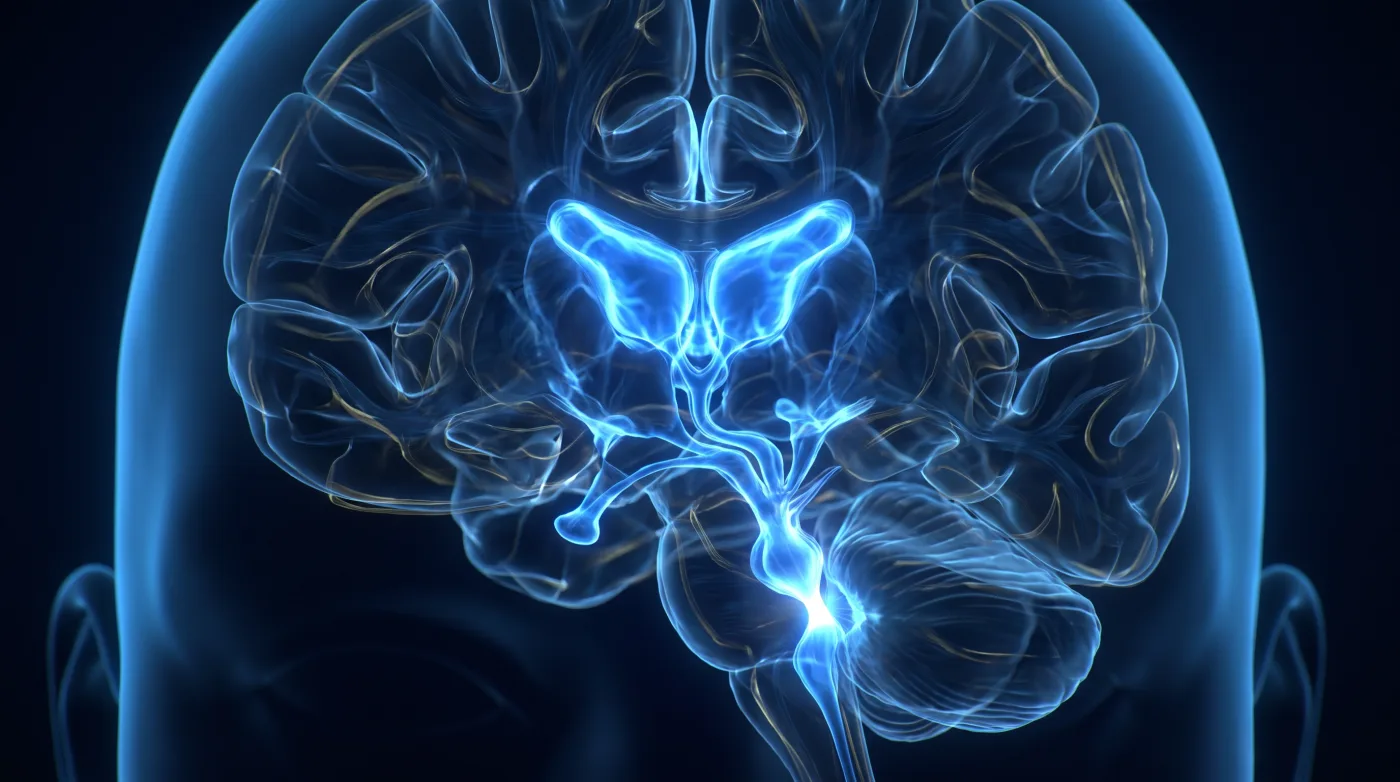
Hydrocephalus: Localize the Block
Big ventricles isn't the question — 'are these ventricles under pressure?' is. Read hydrocephalus as plumbing: find the level of obstruction, then…
Language Mapping: It's the Tracts, Not Just the Cortex
Read language as a dual-stream cortical-plus-white-matter network, not Broca/Wernicke expressive/receptive. Tracts often matter more than cortex…
MR Spectroscopy: The Last Step in the Lesion Tree
No single advanced sequence calls an indeterminate brain lesion — walk a fixed multiparametric tree (enhancement -> diffusion -> perfusion ->…
MRI in Epilepsy: Find the Substrate, Earn the Cure
A 'normal' routine brain MRI in epilepsy is usually a failure of technique and search pattern, not a normal brain. Findings are subtle and finding…
Meningitis: A Normal Scan Doesn't Exclude It
A normal MRI does not exclude meningitis — it's a CSF diagnosis. The scan's real jobs are catching the few specific signs (non-suppressing CSF on…
Multiple Sclerosis: Typical, Changing, or a Red Flag?
Not all white-matter disease is MS, and the MS read isn't 'are there lesions' but 'are they MS-typical, are they changing, and is anything here that…
PML: Subcortical, U-Fiber, and Silent
In an immunocompromised patient, asymmetric confluent subcortical white-matter disease that hugs the U-fibers, spares the overlying cortex, makes…
Post-Treatment Brain MRI: When Enhancement Lies
In the treated brain, enhancement lies in both directions — more isn't always tumor, less isn't always response. Read post-contrast enhancement…
Postoperative Brain Reporting: Answer the Surgeon's Question
On a post-op scan the surgeon already knows what they did — your job is to answer their question, not run a generic search: did anything bleed, is…
Pyogenic Brain Infection: Center Restricts = Abscess
A pyogenic abscess has a stereotyped fingerprint whose load-bearing finding is markedly restricted diffusion in the CENTER of the cavity; center…
Resting-State fMRI: When Task Mapping Fails
Resting-state fMRI exists to rescue the presurgical eloquent-cortex map when task fMRI fails — and it fails often, in exactly the patients you most…
SWI: Where It Blooms and Why
SWI is a single-purpose high-sensitivity detector for things that distort the local field (blood/hemosiderin, calcium, iron, deoxyhemoglobin). Read…
Spontaneous Intracranial Hypotension: The Leak Is in the Spine
The brain findings are the consequence; the leak is in the spine — treat the spinal CSF leak, not the brain. The most dangerous miss is the patient…
Tumor Perfusion: Correct the Leak Before You Trust the Map
Leaky lesions corrupt the CBV map, so before trusting any perfusion number you must know whether leakage was corrected — an uncorrected map can turn…