Brain
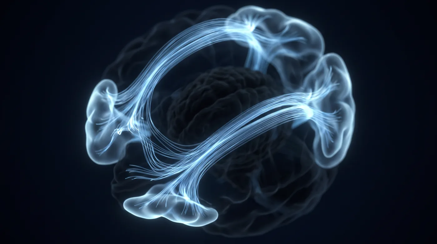
Language Mapping: It's the Tracts, Not Just the Cortex
Fellowship-level neuroradiology teaching by E. Brooke Schrickel, MD. Open this topic in the interactive reading room →
Watch · concise explainerVisual summary
Core clinical idea
Read language as a dual-stream cortical-plus-white-matter network, not Broca/Wernicke expressive/receptive. Tracts often matter more than cortex because cortex reorganizes and white matter does not; a preop language read that doesn't name the at-risk tracts isn't finished.
Bottom line
Language is a dual-stream network — map the tracts, not just the gyri, because cortex reorganizes and white matter doesn't.
Core workstation questions
- Which dominant-hemisphere tracts (arcuate, IFOF, ILF, SLF3, frontal-aslant-type) does this lesion abut or displace, and is the displacement preserving or disrupting them?
- Did this study include DTI? If fMRI-only, is the read incomplete?
- Is my activation phonologic-task or semantic-task driven, and does that explain where it lit up and didn't?
- Do fMRI and WADA (if done) agree on side; if not, am I overcalling dominance?
- Is the posterior frontal activation pars opercularis or ventral premotor cortex — which changes the risk story entirely?
- Have I localized and named SLF3, the arcuate, the IFOF, and the frontal-aslant/striatal tracts relative to the lesion?
- Did I hunt for the anterior basal temporal language area despite susceptibility dropout?
- Did I run phonologic and semantic tasks and read activation against the task used?
What changes reporting / management
- Frame the functional read as what can be safely resected and where the surgeon must stop, not as a list of activations.
- Name the white-matter tracts the lesion abuts; a report localizing only cortex is under-read.
- Mapping changes the operation (improved extent of resection, less OR time, fewer complications, longer survival), so report it as decision-support.
- fMRI gives both lateralization and localization; WADA only lateralization — prefer fMRI as first-line dominance assessment.
- Name pars opercularis vs ventral premotor cortex precisely; ventral premotor is the low-plasticity, high-risk structure for persistent deficit.
- Report SLF3, arcuate, IFOF, and frontal-aslant/striatal tracts by name in dominant-hemisphere preop planning studies.
- Treat the IFOF as the protected ventral semantic tract (it compensates for the ILF/uncinate pathway, not vice versa) and as the anterior limit of temporal lobectomy.
- Use the at-risk tract list to call the resection profile favorable or unfavorable, not just to describe activations.
Practical traps
- Reporting fMRI/WADA concordance you don't have: when they disagree on side, neither is automatically correct.
- Treating fMRI-only mapping as complete — without DTI it under-reads the tract risk that drives outcome.
- Mistaking Heschl's gyrus for language cortex; it processes low-level tones, phoneme work is along the posterior superior temporal sulcus.
- Trusting a negative intraoperative-stimulation map as a safety guarantee; large negative-mapping zones and fMRI-positive/stimulation-negative deficits occur.
- Labeling posterior frontal activation 'Broca's area' when it is ventral premotor cortex — the structure that actually causes lasting speech arrest/deficit.
- Assuming the anterior basal temporal language area is absent because susceptibility dropout obscured it.
- Reading task activation without accounting for whether the task was phonologic or semantic.
- Over-weighting the cortex over a small subcortical/tract lesion that can outweigh it.
Teaching pearls
- Localizing the critical white-matter tracts can matter more than localizing the cortex.
- Heschl's gyrus is tones, not language — phoneme processing lives along the posterior superior temporal sulcus.
- fMRI-only language mapping is an incomplete study; if it lacks DTI, say so.
- A negative stimulation map is not a safety guarantee.
- Language streams are bidirectional — motor-speech machinery is also used in comprehension.
- Ventral premotor cortex — not 'Broca' — is the low-plasticity structure that produces the lasting speech deficit.
- SLF3 and IFOF should be identified in every dominant-hemisphere preop planning study.
- IFOF compensates for the ILF/uncinate pathway, but not the reverse — protect the IFOF.
- Opercular and posterior perisylvian regions are white-matter bottlenecks; one lesion takes several streams.
- Broca's own patient likely deficited from tract (SLF3/frontal aslant) damage, not cortex alone.
Teaching visuals
Source lectures
- Preoperative Language Mapping, Part 1
- Preoperative Language Mapping, Part 2
Educational material for radiology residents and neuroradiology fellows. Nothing here drives individual patient care, and it contains no patient data.