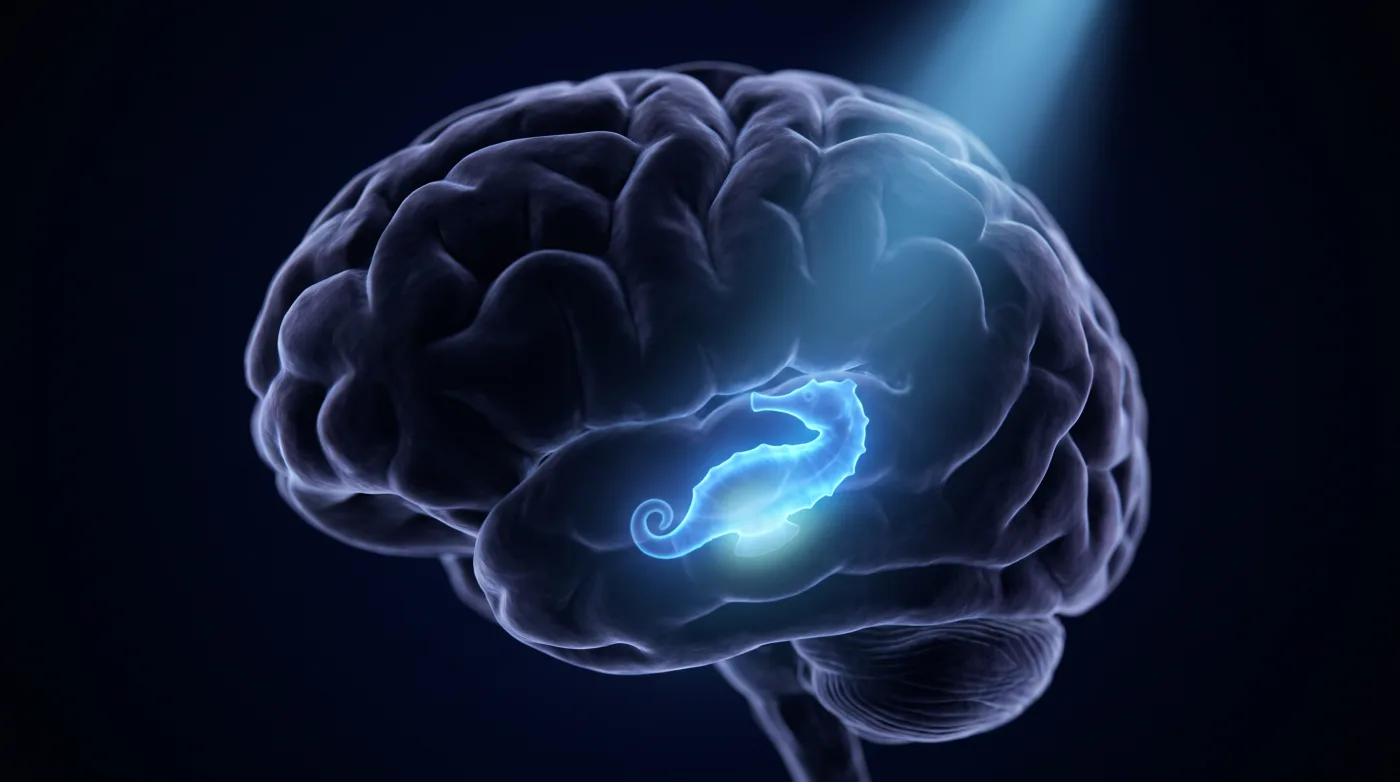
Brain
MRI in Epilepsy: Find the Substrate, Earn the Cure
Fellowship-level neuroradiology teaching by E. Brooke Schrickel, MD. Open this topic in the interactive reading room →
Watch · concise explainerVisual summary
Core clinical idea
A 'normal' routine brain MRI in epilepsy is usually a failure of technique and search pattern, not a normal brain. Findings are subtle and finding the substrate decides whether the patient gets a curative resection — so use a dedicated protocol, run a fixed search (HIPPO SAGE), and know the normal variants cold to neither miss the lesion nor invent one.
Bottom line
Epilepsy MR is a hunt: dedicated protocol, HIPPO SAGE every time, find the substrate, rule out dual pathology, and don't call a variant a lesion.
Core workstation questions
- Is this on a dedicated seizure protocol, and do I need 3T / 3D volumetric sequences for a subtle lesion?
- Is the head rotated (check the IACs) and faking hippocampal asymmetry?
- Is the hippocampus atrophic with increased T2/FLAIR signal and loss of internal architecture?
- Is there a second lesion — dual pathology — that changes the resection?
- Is this cortical thickening / transmantle band a focal cortical dysplasia, and is the true epileptogenic zone wider than the visible abnormality?
- Is this CSF-signal finding just a normal variant (hippocampal sulcus cyst, choroidal fissure cyst)?
What changes reporting / management
- Read on a dedicated seizure protocol; escalate to 3T/3D for developmental or subtle lesions — dedicated protocol plus a trained reader roughly doubles yield over routine brain MRI.
- Suggested phrasing for MTS: hippocampal volume loss with increased T2/FLAIR signal and loss of internal architecture, consistent with mesial temporal sclerosis.
- State when the epileptogenic zone (e.g., in cortical dysplasia) is likely more extensive than the visible MR abnormality — it affects resection margins.
- If truly negative, report MR-negative and recommend a dedicated/3T protocol rather than implying a normal brain.
Practical traps
- Calling a small hippocampus without first excluding head rotation, which causes pseudo-asymmetry.
- Stopping after one finding and missing dual pathology (MTS coexisting with a developmental anomaly); leaving either unresected worsens outcome.
- Mistaking a normal variant (hippocampal sulcus residual cyst or choroidal fissure cyst, both CSF-isointense on all sequences in characteristic locations) for a lesion.
- Confusing focal cortical dysplasia with a low-grade tumor (tumor more often expands/has mass effect) or with heterotopia (periventricular gray matter, not cortical thickening).
Teaching pearls
- An MR-negative epilepsy scan often means wrong protocol or wrong search pattern, not a normal brain.
- Check head rotation before you call a small hippocampus.
- Find one lesion, keep looking — dual pathology changes the operation.
- In cortical dysplasia, the epileptogenic zone is bigger than the blob.
- CSF-isointense on every sequence in a known spot = variant, not pathology.
Teaching visuals
Source lectures
- MR Imaging of Epilepsy
Educational material for radiology residents and neuroradiology fellows. Nothing here drives individual patient care, and it contains no patient data.