Vascular & Stroke
AVM vs Dural AV Fistula: Where the Shunt Lives
Fellowship-level neuroradiology teaching by E. Brooke Schrickel, MD. Open this topic in the interactive reading room →
Watch · concise explainerHow to tell them apart
Core clinical idea
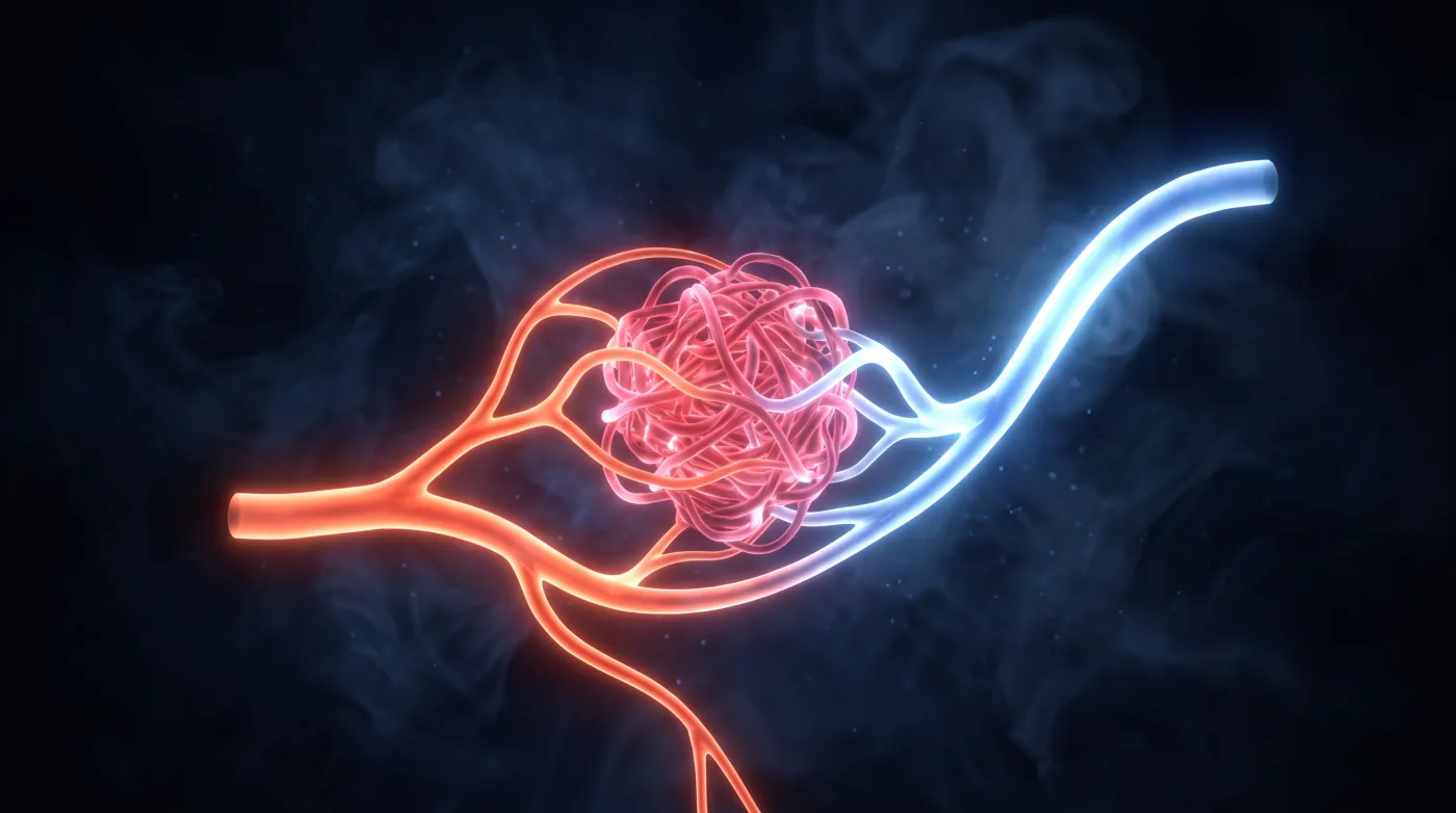
Both are arteriovenous shunts, but where the shunt lives and how it drains decide everything: a true (pial) AVM has a parenchymal nidus fed by cortical/pial arteries, while a dural AV fistula has the shunt inside the dura with NO parenchymal nidus, fed by meningeal/external-carotid branches. In both, the feature that predicts hemorrhage is cortical venous reflux — so the report stands or falls on identifying and grading the venous drainage.
Bottom line
Find the shunt and read its venous drainage — pial nidus (AVM, grade by Spetzler-Martin) vs dural shunt with no nidus (dAVF, grade by cortical venous reflux). Cortical venous drainage is the hemorrhage driver in both.
Core workstation questions
- Is there a parenchymal nidus (AVM) or no nidus at all (dAVF)?
- Who feeds it — pial/cortical arteries (AVM) or dural/meningeal/ECA branches (dAVF)?
- Is there cortical venous reflux / retrograde leptomeningeal venous drainage — the single highest-risk feature?
- For an AVM: what is the size, is it in eloquent cortex, and is venous drainage deep — the three Spetzler-Martin axes?
- Are there flow-related, nidal, or venous (varix) aneurysms that raise rupture risk?
What changes reporting / management
- For an AVM, give the Spetzler-Martin grade: size (<3 cm = 1, 3-6 cm = 2, >6 cm = 3) + eloquent location (+1) + deep venous drainage (+1), total out of 5 — it predicts surgical morbidity.
- For a dAVF, the report hinges on cortical venous reflux: Borden II-III / Cognard IIb and above carry a high hemorrhage/neurologic-deficit risk and warrant treatment; name the involved sinus (transverse-sigmoid, cavernous, tentorial) and location.
- Treat unexplained unilateral cortical venous engorgement, white-matter edema, or venous-pattern hemorrhage as a clue to an occult dAVF and recommend catheter angiography.
- State that DSA remains the reference standard for both — for nidus architecture, feeders, and the venous drainage pattern that grading depends on.
Practical traps
- Missing a dAVF because there is no nidus and the arterial feeders are small — the diagnosis is in the veins (engorged pial veins, venous hypertension).
- Calling dural/ECA arterial supply a pial AVM.
- Under-reporting cortical venous reflux — it is the feature that changes management in both lesions.
- Confusing a benign non-shunting developmental venous anomaly or capillary telangiectasia with an arteriovenous shunt.
Teaching pearls
- AVM = parenchymal nidus with pial feeders; dAVF = dural shunt, no nidus, meningeal/ECA feeders.
- Cortical venous reflux is the hemorrhage driver in both — find it and grade it.
- Spetzler-Martin (size + eloquence + deep drainage) grades AVM surgical risk; Borden/Cognard grade dAVF by venous drainage.
- Unexplained unilateral cortical venous engorgement or edema → hunt for a dural AV fistula.
- A developmental venous anomaly is a normal variant, not a shunt — don't put it in this differential.
Teaching visuals
Source lectures
- AVM vs Dural AV Fistula
Educational material for radiology residents and neuroradiology fellows. Nothing here drives individual patient care, and it contains no patient data.