Spine
Spine Infection: Disc-Centered, Then Hunt the Epidural
Fellowship-level neuroradiology teaching by E. Brooke Schrickel, MD. Open this topic in the interactive reading room →
Watch · concise explainerDisc first, then hunt the epidural
Core clinical idea
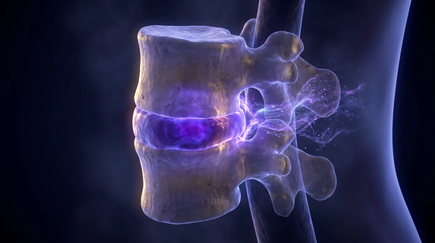
Pyogenic spondylodiscitis is disc-centered: confirm the disc-plus-paired-endplate pattern with endplate erosion, then reflexively hunt the epidural collection compressing the thecal sac — that complication is what turns a medical admission into a surgical emergency. Find the discitis, report the danger.
Bottom line
Confirm disc-plus-paired-endplate infection with erosion, then report the epidural/cord complication — that's the part that changes urgency.
Core workstation questions
- Is this the disc + paired-endplate pattern, and is there true endplate erosion (vs reactive change)?
- Is there an epidural collection compressing the thecal sac — phlegmon or abscess (DWI), where, how much?
- Is the cord signal abnormal (venous congestion/ischemia) — the irreversible complication?
- Is this collection truly epidural, or subligamentous (intact PLL) that I shouldn't upgrade?
- Do the tempo and ESR/CRP fit pyogenic or granulomatous, and does my morphologic read agree?
- Disc destroyed (pyogenic) or disc spared with subligamentous skip lesions and big paravertebral abscess (TB/fungal)?
- Is there endplate (or facet subchondral) erosion — the line between infection and a degenerative/inflammatory mimic?
- Do labs/tempo and plain films/CT agree with infection, or is this tumor (no fever, normal markers, destructive mass) or spondyloarthropathy?
What changes reporting / management
- Commit pattern: T2-bright disc with lost intranuclear cleft, paired endplate edema (T1 dark/T2 bright), endplate erosion, height loss/alignment change, and disc/endplate/paravertebral enhancement.
- Endplate erosion is the discriminator from degenerative mimics — make the erosion call explicitly.
- Exclude epidural extension on every discitis; report presence, location (ventral/dorsal), extent, and degree of thecal-sac compression. Rim-enhancing + DWI-restricting = abscess; homogeneous enhancing tissue = phlegmon.
- Cord T2 signal from epidural venous-plexus congestion/ischemia is a red flag and can be irreversible — escalate, don't sit on it.
- Subligamentous collection (ligaments displaced but PLL intact) is NOT a true epidural abscess — don't upgrade; subdural abscess is rarer, ring-enhancing around the thecal sac, DWI-positive.
- Routes: hematogenous most common, then nosocomial/iatrogenic (Pseudomonas/Klebsiella with instrumentation/catheter/discography/surgery), direct extension, CSF seeding.
- Always compare with CT when available: a vacuum disc phenomenon (intradiscal gas) makes infectious discitis much less likely — don't call the gas 'fluid.' Marrow signal change accompanying a vacuum phenomenon is more often severe reactive/degenerative (Modic) change than infection; weigh the whole pattern before calling discitis-osteomyelitis.
- TB/granulomatous pattern: vertebral signal change with preserved disc spaces, subligamentous anterior/posterior spread, skip lesions, large prevertebral/paravertebral abscess, possible gibbus deformity; fungal can look identical — explicitly search up and down for skip levels.
- Facet septic arthritis = joint fluid PLUS subchondral erosion and perifacet inflammation (can track into the canal behind the thecal sac); fluid alone is not infection.
- Arachnoiditis: nerve-root clumping, peripheral adhesion (empty-sac sign), root/intrathecal enhancement, loculated CSF; chronic disease can calcify (arachnoiditis ossificans).
Practical traps
- Calling intact-PLL subligamentous spread an 'epidural abscess' (overstates surgical picture).
- Missing a true compressive epidural abscess (understates urgency).
- Treating cord signal in this setting as background rather than venous ischemia/infarct.
- Anchoring on disc-destroying pyogenic morphology without checking tempo/ESR/CRP for a granulomatous, disc-sparing pattern.
- Mistaking intradiscal gas (vacuum disc phenomenon) for fluid/infection — vacuum gas argues AGAINST active discitis.
- Demanding disc destruction before calling infection — TB spares the disc, so disc sparing is a feature, not reassurance.
- Calling facet fluid (or disc T2 signal) infection without subchondral/endplate erosion.
- Reading multilevel lytic vertebrae as automatic metastases when coccidioidomycosis or another fungal osteomyelitis fits the host/region.
- Over-calling Modic type 1 reactive endplate change (bright, edematous, enhancing, no erosion) as discitis.
Teaching pearls
- Spondylodiscitis is disc-centered — paired endplate edema plus erosion is the commit pattern.
- Endplate erosion is the line between infection and a degenerative mimic.
- Every discitis read has to work the epidural space — that's where the surgical emergency hides.
- Cord venous congestion/infarct from epidural disease is irreversible — don't sit on it.
- Subligamentous (intact PLL) is not an epidural abscess — don't upgrade it.
- TB spares the disc and skips levels subligamentously — disc sparing is a feature, not reassurance against infection.
- A big paravertebral abscess with preserved discs and skip lesions is TB until proven otherwise.
- Fluid in a facet (or a disc) isn't infection — erosion is.
- No endplate erosion = degenerative (Modic 1), even if it's bright and enhances.
- Multilevel lytic vertebrae can be coccidioidomycosis faking mets; no fever and normal markers point to tumor.
Source lectures
- Spine Infections, Part 1
- Spine Infections, Part 2
Educational material for radiology residents and neuroradiology fellows. Nothing here drives individual patient care, and it contains no patient data.