Spine
Spinal Cord Anatomy: Read It by Column
Fellowship-level neuroradiology teaching by E. Brooke Schrickel, MD. Open this topic in the interactive reading room →
Watch · concise explainerVisual summary
Core clinical idea
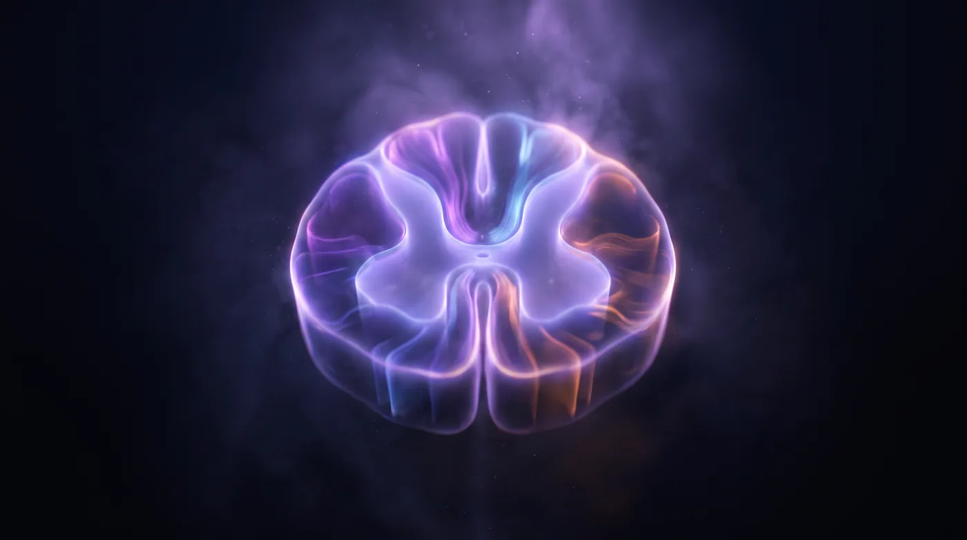
Read the cord by column on the axial image: which column is hit (anterior, lateral, dorsal, central, hemicord) tells you which tracts are down, which names the syndrome, which gives the short differential. Decussation level decides ipsilateral vs contralateral.
Bottom line
Read the cord by column on the axial — the column names the syndrome and the syndrome names the differential.
Core workstation questions
- On axial, which column is involved — anterior, lateral, dorsal, central, or hemicord?
- Does the clinical deficit match that column's tracts (ipsi vs contra by decussation level)?
- Anterior cord + sudden onset: did I look for restricted diffusion (infarct) vs ventral-horn snake-eyes (viral)?
- Dorsal cord: did I name B12 and copper and recommend the labs?
- Saddle anesthesia: is this cauda equina (weakness-dominant) or conus (bladder-dominant), and what is the compressing lesion and level?
What changes reporting / management
- Central cord / dissociated cape-like sensory loss: chase syrinx, Chiari, intramedullary tumor, chronic injury.
- Anterior cord + sudden onset + restricted diffusion = cord infarct (anterior spinal artery) — treat as a vascular event, not nonspecific myelitis; viral/AFM shows ventral-horn snake-eyes.
- Dorsal-column T2 signal is a 'send the labs' finding — name B12 deficiency (vegan diet, nitrous oxide) and copper deficiency (excess zinc, look for peripheral neuropathy).
- Brown-Sequard (hemicord): ipsilateral motor + vibration/position loss, contralateral pain/temp loss (off by a segment or two); trauma classic, lateral demyelinating plaque mimics.
- Cauda equina vs conus is time-critical: name the syndrome, hunt the compressor (epidural abscess, disc/hematoma, conus mass), and report the level; bilateral radicular symptoms can warn of impending cauda equina.
Practical traps
- Reading only the sagittal 'long T2 signal' and skipping the axial column pattern where the diagnosis actually lives.
- Getting ipsilateral vs contralateral backwards by forgetting decussation level (dorsal columns/corticospinal cross high; spinothalamic crosses immediately).
- Sitting on a sudden-onset anterior-cord pattern as 'myelitis' when restricted diffusion makes it an infarct.
- Confusing cauda equina (weakness-dominant) with conus (bladder-dominant) saddle-anesthesia presentations.
Teaching pearls
- Decussation level decides ipsilateral vs contralateral — that's the entire localization trick.
- Dissociated (cape-like) sensory loss sends you to the center of the cord: syrinx, Chiari, tumor.
- Anterior cord + sudden onset + restricted diffusion = infarct, not 'nonspecific myelitis.'
- Dorsal-column signal is a labs finding: B12 (vegan/nitrous) and copper (excess zinc, with neuropathy).
- Saddle anesthesia: cauda equina is weakness-dominant, conus is bladder-dominant — and both want the compressor named.
Teaching visuals
Source lectures
- Functional Anatomy of the Spinal Cord
Educational material for radiology residents and neuroradiology fellows. Nothing here drives individual patient care, and it contains no patient data.