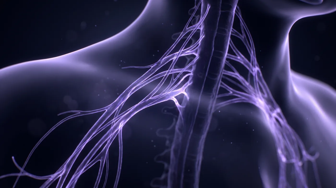
Spine
Brachial Plexus: Preganglionic vs Postganglionic
Fellowship-level neuroradiology teaching by E. Brooke Schrickel, MD. Open this topic in the interactive reading room →
Watch · concise explainerVisual summary
Core clinical idea
The plexus read turns on one divide: preganglionic (root avulsion at the cord — a different surgical problem) vs postganglionic (along trunks/cords/branches). Win it at protocol: cover C4-T2, use 3D T2 to kill CSF pulsation and see rootlets/DRG, key contrast to indication, and don't be fooled by magic angle.
Bottom line
Settle preganglionic vs postganglionic with the right protocol, then localize by fat effacement and denervation pattern — and don't let magic angle fool you.
Core workstation questions
- Is this preganglionic (avulsion: CSF signal, pseudomeningocele, absent rootlets) or postganglionic?
- Did I cover C4-T2, and can I see the rootlets/DRG (3D T2)? If not, is the preganglionic segment non-diagnostic?
- Is the surrounding plexus fat effaced; is there infiltration/displacement?
- What's the muscle denervation pattern, and which level does it localize?
- Is suspicious T2 signal real or magic angle — does it confirm on a second sequence/plane?
- Is there an underlying mass, node, or lung/mediastinal lesion driving this?
What changes reporting / management
- In the report it is often clearer and more clinically useful to localize disease to the SUPRACLAVICULAR vs INFRACLAVICULAR brachial plexus rather than only roots/trunks/divisions/cords — supraclavicular ≈ roots and trunks (at/above the clavicle), infraclavicular ≈ cords and terminal branches (below the clavicle, toward the axilla). It maps to the surgical approach and is easier for the clinician to act on.
- Image C4 through T2 every time to capture prefixed (C4-C8) and postfixed (C6-T2) variants.
- Contrast fork: without contrast for trauma/avulsion and radiculopathy; with contrast for tumor/radiation neuritis, infection, Parsonage-Turner, NF1, polyneuropathy, thoracic outlet.
- Use 3D T2 (SPACE/CUBE) for the preganglionic segment to suppress CSF pulsation and resolve ventral/dorsal rootlets and DRG; coronal T1 + coronal T2 fat-sat (Dixon) for the postganglionic plexus.
- Avulsion = CSF-intensity signal at the foramen, pseudomeningocele, possible contralateral cord displacement, absent rootlets in the pouch; report level and rootlet presence because avulsion vs postganglionic rupture routes the surgery.
- Prioritize C7-T1 in trauma (most-injured, most persistent pain); map muscle denervation (acute edema/enhancement, chronic atrophy/fatty change) back to the level.
- Always sweep for the cause and its company: lymphadenopathy, mediastinal/lung (superior sulcus/Pancoast) mass, incidental neoplasm.
- If rootlets can't be resolved, state the preganglionic segment is non-diagnostic rather than implying normal.
Practical traps
- Magic angle: spurious T2/STIR fat-sat hyperintensity where the plexus runs near ~55 degrees to B0; worst on conventional spin echo, minimized on gradient echo — confirm on a second sequence/plane.
- Narrow FOV clipping a prefixed/postfixed contributing root.
- Tunnel vision on the plexus and missing the underlying mass/node/lung lesion.
- Calling a degraded preganglionic study 'normal' when CSF pulsation obscured the rootlets.
Teaching pearls
- Cover C4-T2 — prefixed and postfixed plexuses will clip a narrow FOV.
- If you can't see the rootlets, you can't exclude avulsion — say so.
- Avulsion vs postganglionic rupture is a different operation — that distinction is the read.
- Fat effacement around the plexus is the tell; denervation pattern localizes the level.
- Plexus signal at ~55 degrees to B0 is magic angle until a second sequence proves otherwise.
- When precise root/trunk/cord localization is hard, describe plexus disease by supraclavicular vs infraclavicular level — it is still clinically actionable.
Teaching visuals
Source lectures
- Practical Brachial Plexus Imaging
Educational material for radiology residents and neuroradiology fellows. Nothing here drives individual patient care, and it contains no patient data.