Pediatrics
Otic Capsule Malformations: How Far Development Got
Fellowship-level neuroradiology teaching by E. Brooke Schrickel, MD. Open this topic in the interactive reading room →
Watch · concise explainerVisual summary
Core clinical idea
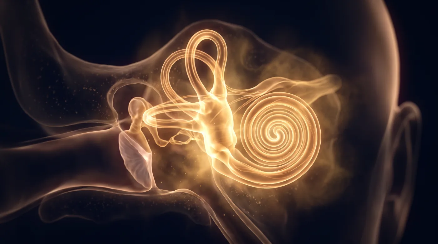
Inner-ear malformations sort by how far cochleovestibular development got before it arrested (Sennaroglu) — so read the cochlea (is it there, does it have a modiolus and ~2.5 turns and interscalar septa), the vestibule and semicircular canals, the vestibular aqueduct, and the cochlear nerve, then translate the pattern into the hearing-rehab option (hearing aid vs cochlear implant vs auditory brainstem implant). The inner ear develops independently of the external/middle ear, so they are usually separate problems.
Bottom line
Grade the developmental arrest (Sennaroglu); check the modiolus, interscalar septa, vestibular aqueduct, and cochlear nerve; and say which rehab the anatomy actually allows.
Core workstation questions
- Is there a cochlea at all — and does it have a normal modiolus and roughly two and a half turns?
- Is the vestibular aqueduct enlarged (the most common imaging malformation, often the only finding)?
- Is the cochlear nerve present on thin T2 (CISS/FIESTA), and is the cochlear aperture / bony canal patent — because a cochlear implant needs a nerve to stimulate?
- Is the modiolus deficient with an enlarged vestibule and EVA (IP-II / Mondini), or absent with the interscalar septa still present (IP-III, X-linked, stapes-gusher risk)?
- Do the cochlea and vestibule form a single undivided cavity (common cavity) versus a separable but cystic cochlea (IP-I)?
- Are the external and middle ear actually normal — they develop independently, so don't assume they are involved (or spared)?
What changes reporting / management
- Name the Sennaroglu type and what it implies for rehab: cochlear implantation needs both a cochlea and a cochlear nerve; cochlear aplasia, common cavity, or cochlear-nerve aplasia point toward an auditory brainstem implant instead.
- Flag IP-III (absent modiolus with preserved interscalar septa, often a bulbous IAC) BEFORE surgery — the defective modiolus/IAC fundus carries a stapes-gusher / CSF-leak risk and an electrode-misplacement risk.
- Report cochlear-nerve aplasia or hypoplasia explicitly — it changes the device and the counseling.
- Identify and flag an enlarged vestibular aqueduct; state the criterion used, and remember EVA can be the only finding and is associated with progressive/fluctuating SNHL.
- Note syndromic associations the pattern suggests (EVA/IP-II with Pendred; IP-III with X-linked deafness; SCC aplasia with CHARGE; apical cochlear hypoplasia with branchio-oto-renal) so the right work-up follows.
Practical traps
- Calling a common cavity an incomplete partition (IP-I has a separable, if cystic, cochlea and vestibule; a common cavity is one undivided sac).
- Promising a cochlear implant without confirming the cochlear nerve on thin T2 — an absent nerve changes the operation to an ABI.
- Forgetting that the enlarged vestibular aqueduct is the commonest malformation and may be the only clue — scan for it deliberately.
- Assuming the external/middle ear is involved because the inner ear is abnormal; they develop separately and concomitant involvement is unusual.
- Over-reading subtle asymmetry as malformation without the dedicated thin-section CT and high-resolution T2 to prove it.
Teaching pearls
- IP-II is the classic Mondini triad: an apical modiolar defect, a mildly dilated vestibule, and an enlarged vestibular aqueduct — and it is the most common cochlear malformation.
- IP-III = absent modiolus but the interscalar septa are still present — think X-linked deafness and warn about the stapes gusher.
- Cochlear implant needs a cochlea AND a cochlear nerve; without one, the answer is an auditory brainstem implant.
- The vestibular aqueduct is enlarged when it is bigger than the adjacent posterior semicircular canal — an easy at-the-scanner check.
- Inner ear vs external/middle ear develop independently — read them as separate questions.
Teaching visuals
Source lectures
- Otic Capsule & Inner Ear Malformations
Educational material for radiology residents and neuroradiology fellows. Nothing here drives individual patient care, and it contains no patient data.