Head & Neck
Craniocervical Junction Trauma: Ligaments, Not Just Bones
Fellowship-level neuroradiology teaching by E. Brooke Schrickel, MD. Open this topic in the interactive reading room →
Watch · concise explainerVisual summary
Core clinical idea
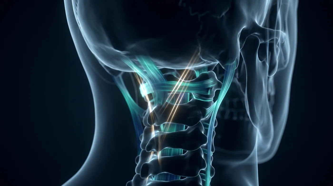
At the craniocervical junction the grave, potentially fatal injury is often purely ligamentous with no fracture — because the CCJ is held by ligaments, not bony interlock. Don't clear it on bones alone: hunt malalignment and abnormal distances, and chase the associated cord and vascular injuries.
Bottom line
At the craniocervical junction the grave injury is often purely ligamentous with no fracture — clear it on alignment and distances, not bones, and chase the associated cord and vascular injury.
Core workstation questions
- Have I cleared this on alignment and distances, or only on 'no fracture'?
- Are the key distances increased — basion–dens, condyle–to–C1 lateral mass, atlanto-dental interval?
- If I found AOD, did I measure for atlanto-axial dislocation too (they coexist)?
- Did I check coronals for the medial condyle avulsion and the odontoid base fracture?
- Given any CCJ ligamentous concern, have I prompted MRI for cord/ligaments and imaging for vertebral artery injury?
- Is this asymmetric dens–lateral-mass distance real, or just head rotation — and is it supported by any other sign?
What changes reporting / management
- Do not clear the CCJ on absence of fracture; report instability when distances/alignment are abnormal even with intact bone.
- AOD signs: increased basion–dens distance, increased skull-base-to-C1 posterior arch, increased occipital condyle–to–C1 lateral mass (atlanto-occipital interval).
- Atlanto-axial dislocation signs: increased atlanto-dental interval, transverse ligament disruption, abnormal C1–C2 signal/edema.
- Prompt MRI for ligaments and cord, and vascular imaging for vertebral artery injury, when CCJ ligamentous disruption is suspected.
- Find one dislocation pattern, deliberately measure for the other — AOD and atlanto-axial dislocation coexist.
Practical traps
- Clearing the CCJ on 'no fracture' and missing a purely ligamentous, unstable injury.
- Missing the medial occipital condyle avulsion on axials (check coronals).
- Over-calling asymmetric dens–lateral-mass distance caused by head rotation when unsupported by other signs.
- Stopping at the osseous classification without evaluating distances, ligaments, cord, and vessels.
Teaching pearls
- No fracture does not clear the CCJ — the lethal injury can be purely ligamentous.
- Increased basion–dens distance should make you think atlanto-occipital dislocation.
- AOD and atlanto-axial dislocation travel together — find one, measure for the other.
- The medial occipital condyle avulsion hides on axials — check the coronals.
- Asymmetric dens–lateral-mass distance is a soft sign — head rotation fakes it; trust it only in company.
Teaching visuals
Source lectures
- Evaluation of CCJ Trauma
Educational material for radiology residents and neuroradiology fellows. Nothing here drives individual patient care, and it contains no patient data.