Head & Neck
Adult Cystic Neck Mass: Cancer Until Proven Otherwise
Fellowship-level neuroradiology teaching by E. Brooke Schrickel, MD. Open this topic in the interactive reading room →
Watch · concise explainerVisual summary
Core clinical idea
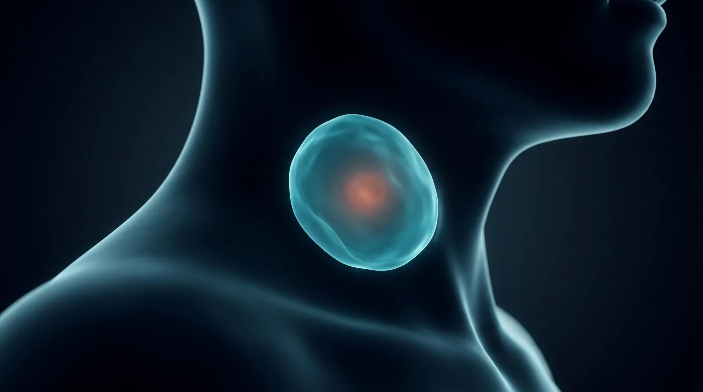
A cystic neck mass in an adult is squamous cell carcinoma (or cystic papillary thyroid met) until proven otherwise — never default to branchial cleft cyst; the most reassuring-looking cysts are exactly the metastatic nodes.
Bottom line
A cystic neck mass in an adult is squamous cell carcinoma until proven otherwise — diagnose developmental cysts by their confirming feature, recognize mimics by location, and never default to branchial cleft cyst.
Core workstation questions
- Is this a node? If cystic and nodal in an adult, am I working it up as cancer?
- For a cystic node, where's the primary — thyroid bed/TE groove (papillary thyroid) or tonsil/tongue base (HPV-SCC)? Any piriform sinus expansion?
- For a presumed developmental cyst, do I see the confirming feature (sublingual communication for ranula, true midline for Thornwaldt)?
- Could this low-density node be TB or atypical mycobacterial rather than necrotic SCC?
- Is this benign mimic in its characteristic location (thoracic duct at venous confluence, parathyroid cyst elevating thyroid lobe), or am I over-calling a variant?
What changes reporting / management
- Treat an adult cystic node as malignant: recommend primary-tumor search and tissue diagnosis rather than labeling a benign developmental cyst.
- Cystic node with eccentric/absent mural nodularity and thin wall favors papillary thyroid met; thick viable rind around necrosis favors SCC nodal met.
- Postoperative neck fluid: seroma resolves in ~2 weeks; persistent collection after thoracic duct injury is a chyloma needing surgical correction.
- Off-midline nasopharyngeal cyst is a retention cyst, not Thornwaldt; report accordingly.
Practical traps
- Calling a smooth, thin-walled cyst anteromedial to the SCM a branchial cleft cyst in an adult (it is the textbook look of a metastatic cystic node).
- Being reassured by a benign-appearing cystic node and not searching for a primary.
- Mistaking low-density TB/atypical mycobacterial nodes for necrotic SCC, or vice versa.
- Escalating a chronically inflamed/old infected ranula to malignancy when the sublingual communication is present.
- Mistaking a dilated thoracic duct at the venous confluence for pathology.
Teaching pearls
- In an adult, it's almost never a branchial cleft cyst — assume squamous cell carcinoma.
- A purely cystic node sends you hunting: thyroid bed and TE groove for papillary thyroid, tonsil and tongue base for HPV-SCC.
- Ranula is confirmed by communication with the sublingual space; Thornwaldt by true midline location.
- Thoracic duct at the IJ–subclavian confluence is normal — recognize the location before calling it pathology.
Teaching visuals
Source lectures
- Adult Cystic Neck Masses
Educational material for radiology residents and neuroradiology fellows. Nothing here drives individual patient care, and it contains no patient data.