Head & Neck
Sinonasal Maldevelopmental Lesions: One Event, One Family
Fellowship-level neuroradiology teaching by E. Brooke Schrickel, MD. Open this topic in the interactive reading room →
Watch · concise explainerVisual summary
Core clinical idea
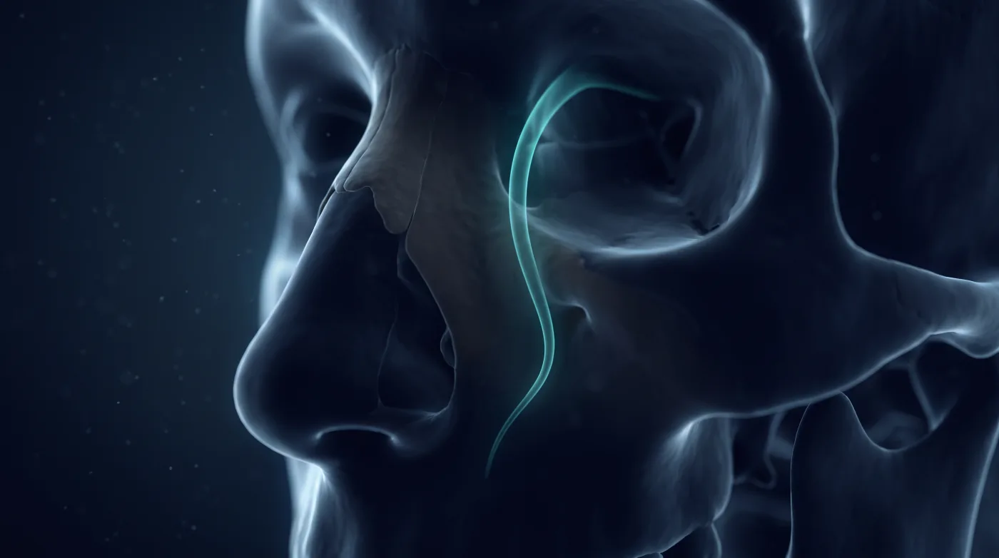
One developmental event explains the whole family: a dural diverticulum dips into the prenasal space behind the nasal bone and normally regresses (leaving the foramen cecum). Persist = encephalocele, pinch off = glial heterotopia, drag ectoderm back = dermal sinus/dermoid. All are cranionasal, so any superior nasal mass abutting the anterior fossa must have its cribriform/fovea ethmoidalis cleared before it is called benign.
Bottom line
One dural diverticulum explains encephalocele, glial heterotopia, and dermal sinus — and every superior nasal mass gets its anterior fossa floor cleared before it's called benign.
Core workstation questions
- Is the cribriform/fovea ethmoidalis intact, or is this superior nasal mass continuous with an anterior fossa defect?
- Which mechanism fits — herniating CNS (encephalocele), pinched-off CNS over the dorsum (glial heterotopia), or a tract/dermoid to the foramen cecum (dermal sinus)?
- For an encephalocele, what are the roof and floor (naming), and is this the sneaky intranasal nasoethmoidal one?
- For a dermal sinus, how far intracranially does the tract go (full-resection planning)?
- Sphenoid wing dysplasia — are there other NF1 features, and is a small greater-wing defect just benign fat herniation rather than tumor/trauma?
What changes reporting / management
- Prove the cribriform plate/fovea ethmoidalis is intact on any superior nasal mass; a defect raises encephalocele and should stop blind transnasal biopsy/resection.
- Name encephaloceles roof-first/floor-second (frontonasal, nasoethmoidal) and specifically flag the adult intranasal nasoethmoidal encephalocele masquerading as a polyp.
- Trace a nasal dermal sinus/dermoid to the foramen cecum and report full intracranial extent for complete resection.
- Recognize sphenoid wing dysplasia (empty orbit, temporal lobe/subarachnoid herniation, pulsatile proptosis), link to NF1, and don't mistake a small benign greater-wing fat-herniation defect for tumor/trauma.
Practical traps
- Calling herniated or heterotopic CNS tissue a polyp and biopsying it.
- Missing the intranasal nasoethmoidal encephalocele in an adult superior nasal mass.
- Labeling glial heterotopia a 'nasal glioma'/neoplasm (it is GFAP/S100-positive heterotopic glial tissue, often gliotic).
- Confusing a small greater-wing fat-herniation defect with post-traumatic or neoplastic disease.
Teaching pearls
- Persist = encephalocele, pinch off = glial heterotopia, drag ectoderm = dermal sinus — one mechanism, three lesions.
- The adult intranasal nasoethmoidal encephalocele is the polyp that's actually brain — check the cribriform plate.
- 'Nasal glioma' is heterotopic glial tissue (GFAP/S100+), not a tumor.
- Resect the nasal dermal sinus tract in full to the foramen cecum, or it recurs.
- Sphenoid wing dysplasia (empty orbit, pulsatile proptosis) — think NF1, and don't confuse a fat-herniation defect for tumor.
Teaching visuals
Source lectures
- Sinonasal Maldevelopmental Lesions
Educational material for radiology residents and neuroradiology fellows. Nothing here drives individual patient care, and it contains no patient data.