Head & Neck
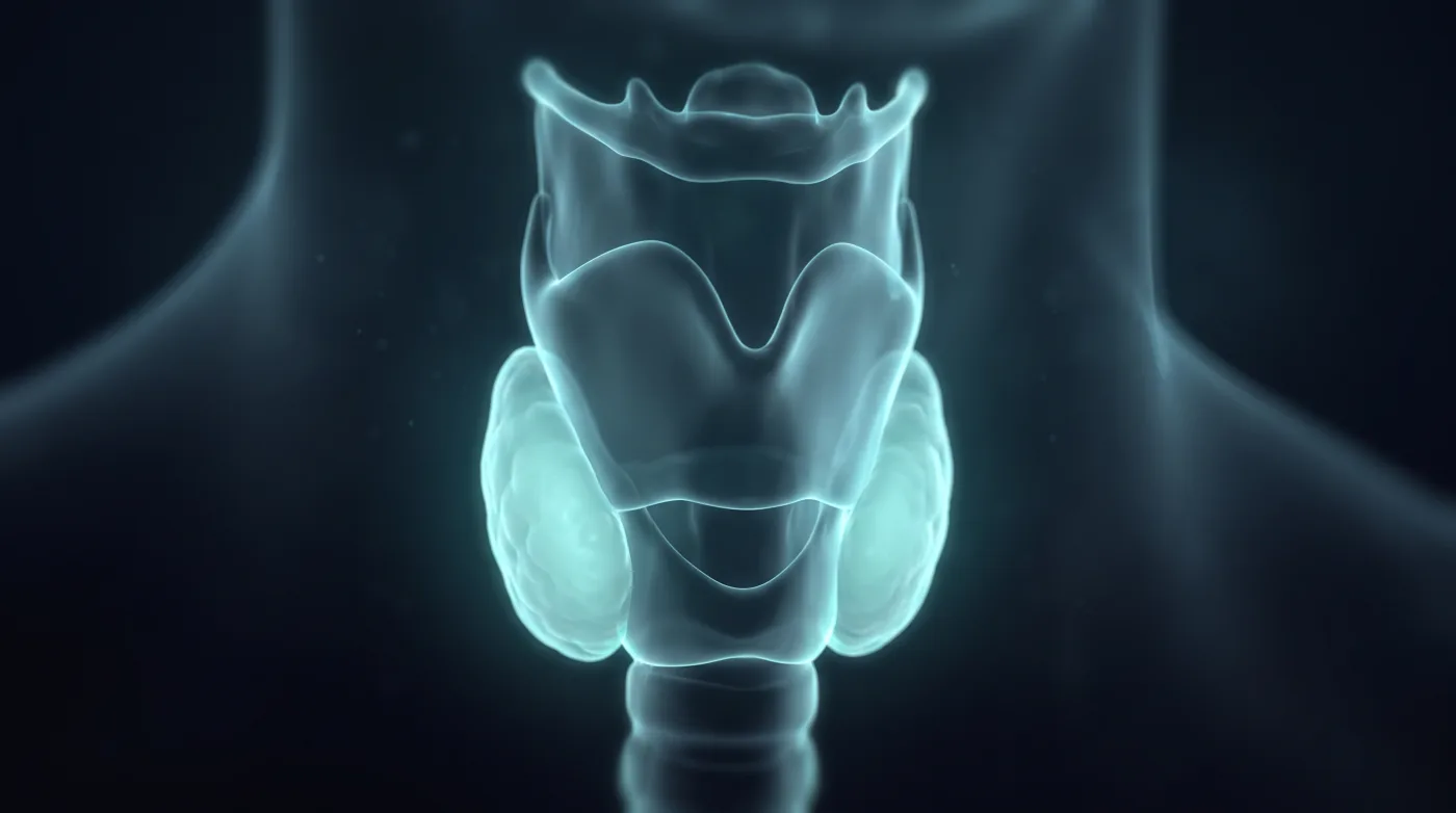
Larynx: The Two Fat Spaces That Upstage It
Fellowship-level neuroradiology teaching by E. Brooke Schrickel, MD. Open this topic in the interactive reading room →
Watch · concise explainerVisual summary
Core clinical idea
Learn larynx anatomy because two fat spaces and one cartilage set the stage: effacement of the pre-epiglottic or paraglottic fat upstages to at least T3, and through-and-through thyroid cartilage penetration upstages to T4a. Read every laryngeal CT as subsite + fat space + cartilage; your value over the scope is the submucosal extent the surgeon can't see.
Bottom line
Larynx anatomy is a staging tool — two fat spaces make it T3, the thyroid cartilage makes it T4a, and MRI settles the cartilage.
Core workstation questions
- What subsite is this — where is the laryngeal ventricle and which structures are involved (supraglottis / glottis / subglottis)?
- Is the pre-epiglottic or paraglottic fat effaced? (At least T3.)
- Is there through-and-through thyroid cartilage penetration? (T4a — and is the cartilage ossified enough to judge on CT?)
- Should this go to MRI because the cartilage call would change definitive chemoradiation vs laryngectomy?
- Is the AE fold / pyriform region the real primary, i.e. larynx vs hypopharynx?
- What does the clinical note say about mucosal extent and cord mobility? (Staging needs it.)
- Cartilage: just sclerosis (coin flip) or true through-and-through penetration (T4a)? Does the answer change surgery, and are the cords already fixed?
- Is there strap-muscle/extralaryngeal spread (T4a) or prevertebral/carotid involvement (T4b, unresectable)?
What changes reporting / management
- Call effacement of the pre-epiglottic or paraglottic fat as at least T3 disease; use sagittal MR for the cleanest pre-epiglottic fat read.
- Report through-and-through thyroid cartilage penetration as T4a, recognizing it can shift the patient from voice-sparing chemoradiation to laryngectomy.
- Recommend MRI as the cartilage-involvement problem-solver when the CT cartilage call would change management; CT is otherwise the staging workhorse.
- Identify the subsite off the false cord / ventricle / true cord stack and the three-cartilage true-cord level before assigning extent.
- Call pre-epiglottic or paraglottic fat effacement as at least T3 without needing clinical cord-fixation history; report through-and-through thyroid cartilage as T4a and strap-muscle/extralaryngeal tumor as T4a.
- Treat CT cartilage sclerosis as nondiagnostic (~50/50 tumor vs perichondritis); escalate to MRI only when the cartilage answer changes surgery and the cords aren't already fixed.
- On MRI, require tumor-matching signal on T1, T2, and post-contrast for cartilage invasion; T2 brighter than tumor indicates edema/perichondritis, not tumor.
- Answer the surgeon's four questions every time: subsite(s); extralaryngeal/hypopharynx involvement; pre/paraglottic fat and cartilage; nodes (with bilaterality for supraglottic).
Practical traps
- Calling cartilage invasion on CT when the thyroid cartilage is non-ossified or irregularly ossified and there's no through-and-through strap involvement.
- Reporting only mucosal-level findings the scope already showed, while missing the submucosal fat-space and cartilage extent.
- Mistaking an axial slice through the cricothyroid membrane (posterior cricoid present, no anterior cartilage) for a cartilage defect.
- Confusing an AE-fold/pyriform tumor's primary site — larynx vs hypopharynx.
- Dictating 'likely cancer' or an imaging-only T-stage when staging depends on clinical cord mobility.
- Over-calling cartilage invasion from CT sclerosis alone (asymmetric arytenoid sclerosis occurs in normals).
- Recommending a cartilage MRI without checking cord mobility first (a fixed larynx may go to laryngectomy regardless).
- Misassigning the primary site of a large AE-fold/pyriform mass (larynx vs hypopharynx).
Teaching pearls
- Pre-epiglottic or paraglottic fat effaced = at least T3.
- Through-and-through thyroid cartilage = T4a — and that flips chemoradiation to laryngectomy.
- Non-ossified cartilage is the CT trap; that's the MRI case.
- Find the ventricle and you've found the subsite map.
- No anterior cartilage on an axial cut through the cricoid is the cricothyroid membrane, not a defect.
- 'Likely cancer' is not a staging report — give submucosal extent.
- Pre/paraglottic fat = T3; through-and-through thyroid cartilage = T4a.
- CT cartilage sclerosis is 50/50 — sclerosis alone isn't invasion.
- MRI cartilage call needs tumor-matching signal on T1, T2, and post-contrast; T2 brighter than tumor is edema.
- Strap-muscle tumor is extralaryngeal spread — that's T4a.
Teaching visuals
Source lectures
- Larynx: Imaging Anatomy
- Larynx: SCCA & Pathways of Spread
Educational material for radiology residents and neuroradiology fellows. Nothing here drives individual patient care, and it contains no patient data.