Head & Neck
Head & Neck Cancer: The Fat Planes That Stage It
Fellowship-level neuroradiology teaching by E. Brooke Schrickel, MD. Open this topic in the interactive reading room →
Watch · concise explainerVisual summary
Core clinical idea
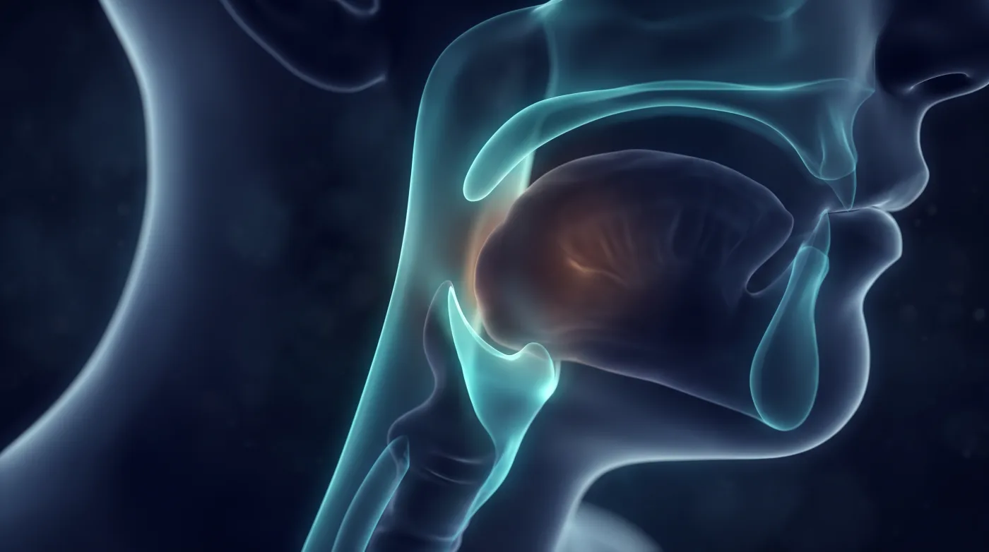
Learn the oropharynx as the boundaries and fat planes that staging turns on, not as a muscle tour: correct subsite assignment (tongue base = oropharynx, oral tongue = oral cavity, epiglottis = larynx), the retropharyngeal/prevertebral and parapharyngeal fat planes for deep invasion, and the carotid relationship that decides operability.
Bottom line
Read oropharynx anatomy for staging — nail the subsite boundaries, interrogate the retropharyngeal and parapharyngeal fat for deep invasion, and flag the carotid relationship that decides operability.
Core workstation questions
- Which subsite is this, and is the boundary right (circumvallate papillae for tongue base vs oral tongue; epiglottis is larynx)?
- Is the retropharyngeal/prevertebral fat plane preserved or effaced — and am I on MR if it's too thin to judge on CT?
- Is the parapharyngeal fat clean, or is tumor extending into it?
- Where is the internal carotid relative to a tonsillar tumor — does this take transoral robotic surgery off the table?
- Did I check the mandibular and vertebral marrow fat for invasion?
- Does this node have a fatty hilum and homogeneous enhancement, or necrosis/heterogeneity? (Architecture, not size.)
- Is the perinodal fat preserved, or is there extracapsular spread into adjacent muscle or gland?
- What AJCC level is it, assigned by landmark — and is that the word the surgeon will use?
What changes reporting / management
- Upstaging anatomy is the report's job — name the specific structure that pushes a primary to T3/T4. Larynx/hypopharynx: vocal-cord FIXATION and pre-epiglottic/paraglottic fat invasion = T3; inner-cortex thyroid cartilage = T3, and through-cartilage or extralaryngeal spread (strap muscles, trachea, esophagus, thyroid gland, soft tissues of the neck) = T4a.
- Oral cavity: cortical mandible/maxilla, inferior alveolar canal, extrinsic tongue muscles, maxillary sinus, or facial skin = T4a; masticator space / pterygoid plates, skull base, or internal carotid encasement = T4b. Nasopharynx: skull-base bone, pterygoid, paranasal sinuses, or cervical vertebra = T3; intracranial extension, cranial nerves, orbit, hypopharynx, parotid, or beyond the lateral pterygoid = T4.
- Sinonasal (maxillary): orbital floor/medial wall, pterygoid fossa, or ethmoid = T3; anterior orbit, cheek skin, cribriform plate, pterygoid plates, or infratemporal fossa = T4a; orbital apex, dura/brain, middle cranial fossa, clivus, nasopharynx, or ICA = T4b. For every primary, ask which adjacent structure — if invaded — makes this T3/T4, and report it explicitly.
- Assign the correct subsite (base of tongue, soft palate, tonsillar region, posterior wall); tongue base is oropharynx, epiglottis is larynx.
- Report status of the retropharyngeal/prevertebral fat plane (preservation argues against deep/prevertebral fascia invasion).
- Report parapharyngeal fat involvement as a sign of deep spread.
- Note internal carotid position relative to a tonsillar tumor — abutment/posterior position can contraindicate transoral robotic surgery.
- Use non-fat-suppressed T1 for overall anatomy; contrast/fat-sat sequences for problem-solving and lesion definition.
- Describe necrosis/heterogeneity and a lost fatty hilum explicitly; do not pass a node on size alone if the architecture is abnormal.
- Report extracapsular spread (lost perinodal fat plane, infiltrative margin into muscle/gland) — it is an AJCC staging modifier, not descriptive color.
Practical traps
- Assigning an epiglottic finding to the oropharynx (it is larynx).
- Miscalling tongue base vs oral tongue by missing the circumvallate-papillae divide.
- Calling an off-center uvula a mass (more of a pitfall on CT).
- Failing to evaluate the fat planes for deep invasion because they are thin on CT.
- Innervation trap: of the extrinsic tongue muscles, only palatoglossus is CN X (pharyngeal plexus); genioglossus/hyoglossus/styloglossus are CN XII.
- Equating sub-centimeter with benign — a large share of pathologically positive nodes are under a centimeter.
- Reporting a node without its AJCC level or without commenting on the capsule.
- Missing retropharyngeal, level VII, and TE-groove nodes that sit outside the obvious chains.
- Calling a Virchow (left supraclavicular) node a head-and-neck node when it points at a chest/abdominal primary.
Teaching pearls
- For every H&N primary the upstaging question drives the read: which adjacent structure, if invaded, makes this T3 or T4 — cartilage, cord fixation, deep extrinsic muscle, bone, skull base, or carotid? Name it.
- Tongue base is oropharynx, oral tongue is oral cavity — the circumvallate papillae are the line.
- The epiglottis sticks up into the oropharynx but it's larynx — don't assign its findings here.
- Two fat planes decide deep invasion: retropharyngeal/prevertebral and parapharyngeal — look at both, often on MR.
- Note the carotid relative to a tonsillar tumor — it can contraindicate transoral robotic surgery.
- Sub-centimeter is not benign — tumor-packed nodes are often normal-sized.
- Necrosis or a lost fatty hilum beats any size threshold.
- Extracapsular spread is a staging variable — call it, don't just imply it.
- Speak the surgeon's levels, or the report won't drive the dissection.
- A node in the wrong drainage territory means look harder for a different primary.
Teaching visuals
Source lectures
- Head & Neck Cancer - The Oropharynx Part 1: Anatomy
- Head and Neck Cancer: Lymphadenopathy
Educational material for radiology residents and neuroradiology fellows. Nothing here drives individual patient care, and it contains no patient data.